news
Brain MRI predicts early dementia progression
New research in *Neurology* suggests that specific brain atrophy patterns seen on MRI scans can predict how fast early-onset dementia progresses. Researchers, led by Alexandra Touroutoglou from Harvard Medical School, found that atrophy in key areas associated with early Alzheimer's helps forecast the timeline of cognitive decline. This could mean earlier, more precise insights into how someone's brain health will change.
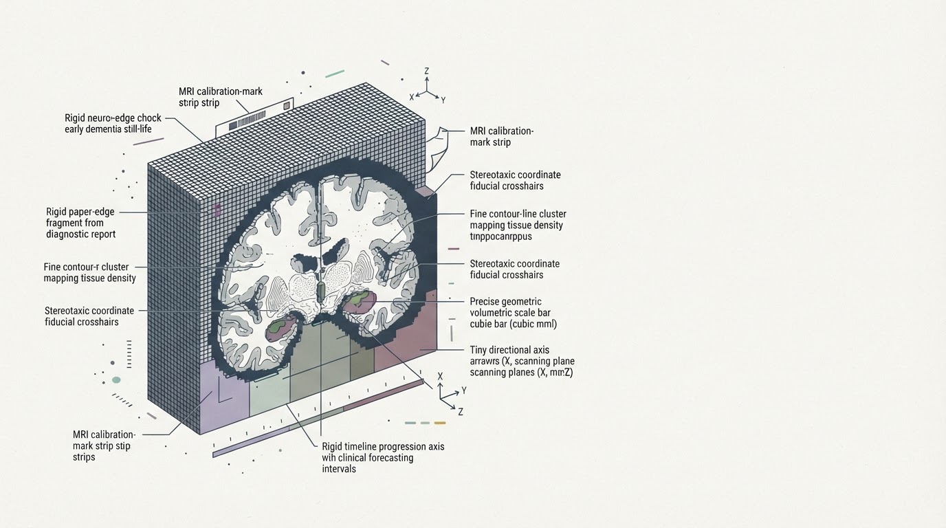
New research in Neurology suggests that specific brain atrophy patterns seen on MRI scans can predict how fast early-onset dementia progresses. Researchers, led by Alexandra Touroutoglou from Harvard Medical School, found that atrophy in key areas associated with early Alzheimer's helps forecast the timeline of cognitive decline. This could mean earlier, more precise insights into how someone's brain health will change.
The architecture take
I’ve always said that brain scans will eventually tell us more than memory tests alone, and here's a step in that direction. This new research points to specific areas of the brain where atrophy—which is just a fancy way of saying tissue loss—can actually predict how quickly early-onset dementia will progress. It's not just a general shrinkage; it’s targeted. The team, led by Dr. Alexandra Touroutoglou, zeroed in on regions already known to be affected in early Alzheimer's. This isn't about diagnosing the disease, but about giving doctors a clearer picture of its timeline once it's already there. For someone living with mild cognitive impairment, knowing whether their brain is deteriorating slowly or quickly could change everything about how they plan their life and treatment. It also makes me wonder what preventative measures could slow down that specific atrophy, long before it becomes a problem.